Answer a specific question
Imaging is selected to support a clinical decision. Your dentist might investigate a symptom, evaluate a suspected cavity, or review an area before planning treatment.


Explore your care
Everyday care, tooth repair and support for your gums.
Explore your care
Explore the possibilities for your smile.
Explore your care
Options for replacing one tooth, several teeth or a full arch.
Explore your care
Explore tooth extractions and wisdom teeth removal.
Explore your care
Explore treatment options for the inside of your tooth.
Explore your care
Two ways to explore a straighter smile.
Atlas Dental
Atlas Dental
General Dentistry / Treatment guide
Dental X-rays provide information about areas your dentist cannot see directly. The type of image depends on the question being investigated, such as decay between teeth, a tooth root, or the bone supporting your smile.
Explore your options with our Atlas Dental team. Ask who would provide your treatment and whether a specialist referral is needed.
Let’s talk about your options See what care can involve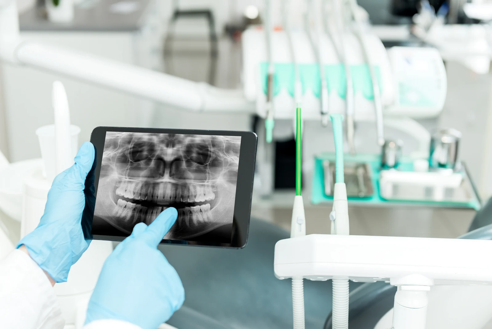
A panoramic X-ray gives the dentist a broad view of the teeth, roots and jaws.
This is one type of dental X-ray. Smaller views may be needed to answer a specific question about a tooth or its supporting bone.
Understanding your care
Start with the purpose of treatment, then explore what matters for your own care.
Imaging is selected to support a clinical decision. Your dentist might investigate a symptom, evaluate a suspected cavity, or review an area before planning treatment.
Previous images may provide a useful reference. Sharing available records can help your dentist understand your history and decide whether new images are needed now.
Small intraoral images and wider views provide different information. More complex imaging is considered when its additional detail is relevant to the problem being assessed.
From questions to a plan
The details and sequence depend on your assessment and individual treatment plan.
Your dentist reviews your history, symptoms, and examination to determine whether imaging is likely to provide useful information for diagnosis or treatment planning.
The team positions you and the imaging equipment for the selected view. A small sensor may be placed inside your mouth for some images.
You remain still while the image is taken. The team checks that the area needed is visible before deciding whether any other views are justified.
The dentist interprets the image with the rest of the examination. Ask to see the area being discussed and how it affects your treatment options.
Before you decide
Dental X-rays use ionizing radiation, so images should be justified by a clinical need. Modern techniques limit exposure, but additional imaging is not automatically better. Images also have limitations and must be interpreted alongside an examination. Tell your team about your health circumstances and any difficulty tolerating sensors so they can plan appropriately.
There is no single interval for everyone. Age, symptoms, existing disease, risk factors, and earlier images all influence when another view is likely to be useful.
Often, previous images are useful for comparison or planning. Their age, quality, and coverage affect whether they answer the current question or whether another image is needed.
No. An image adds information about hidden structures, while the examination provides information about symptoms, tooth surfaces, gums, and soft tissues that an image alone cannot establish.
A place to begin
Talk through your priorities, alternatives and who would provide your care. An assessment helps clarify the next step.
Let’s talk about your optionsOur team will contact you to confirm your visit.
The treatment images and animations in this guide illustrate examples of care. They are not Atlas Dental patient results, personal treatment recommendations or statements about an individual clinician’s scope. Your assessment and care plan determine which options are appropriate for you.